More about HKUST
(This article was originally published on EurekAlert! on December 14, 2023)
A research team at the Hong Kong University of Science and Technology (HKUST) has outlined the high-resolution structure of a little-known virus, improving our understanding of viral infection, which could pave the way for more accurate predictions of climate change.
With the help of an advanced technique involving cryo-electron microscopy, they managed to capture images of the virus – the cyanophage P-SCSP1u – at near-atomic resolution in its native form and examined it to see how its different parts fit together. This helped show how different proteins work in the virus and how they interact to make the virus stable and able to infect cells.
Cyanophages, together with their host cyanobacteria, play important roles in marine biogeochemical cycles and control of marine food webs. P-SCSP1u is a representative member of a group of cyanophages called MPP-C, which contain the smallest genomes among cyanopodoviruses and act differently when they infect bacteria. But little is known about their infection process given the lack of high-resolution structural information.
The team, led by Prof. DANG Shangyu, Assistant Professor of the Division of Life Science, and Prof. ZENG Qinglu, Associate Professor of the Department of Ocean Science and the Division of Life Science at HKUST, used an advanced technique – the single-particle cryo-electron microscopy (cryo-EM) – to examine the virus up close, revealing the assembly mechanism of the head and molecular interaction of a part of the cyanophage called the portal-tail complex.
By comparing it to other similar viruses with known structures, the team gained insights into how different components of these viruses have changed over time during the infection process.
Additionally, the study gives us a close-up look at the portal-tail complex, which helps control the virus's DNA. By comparing it to a similar structure in a different virus called phage T7, the study shows how these types of viruses control their DNA. The team also found out about two new parts – a valve and a gate – that help these viruses control how they let DNA in and out of themselves.
“We believe this high-resolution native structure of cyanophage is an important and timely contribution to the field of bacteriophages for the understanding of viral infection,” said Prof. Dang.
Cyanophages are viruses that infect cyanobacteria, which are photosynthetic bacteria that play a crucial role in the Earth's ecosystems. Besides oxygen, they also provide a lot of the food and fuel we consume in our daily lives. These viruses are specific to infect cyanobacteria, and they can significantly impact the population and behavior of these bacteria. These are some of the reasons why understanding how these bacteria grow and die is critical for figuring out how much carbon is in the oceans.
In sunny water, about 5 percent of these cyanobacteria are infected by cyanophages. When infected, these viruses take over the bacteria and make them produce more viruses, akin to turning the bacteria into a virus-producing factory.
These viruses can affect how the bacteria make energy from sunlight by stopping them from using CO2, causing a loss of a huge amount of carbon each year, more than what all the coral reefs, marshes, and sea plants on Earth produce, Prof. Zeng explained.
Figuring out how these viruses work and infect their hosts is crucial, as it helps scientists understand how they affect the bacteria, the oceans, and even the climate, he said.
“Cyanobacteria are highly abundant in Hong Kong waters and play a major role in global CO2 fixation. The global abundance and carbon fixation capacity of cyanobacteria are important for calculating the marine carbon budget. Cyanophages are a major cause of cyanobacteria death. Studying the infection process of cyanophages is important for understanding how they regulate cyanobacterial populations, which is crucial for the accurate estimation of the global carbon budget and for the prediction of climate change,” Prof. Zeng added.
Understanding how these viruses infect can help facilitate the future use of viruses to control harmful cyanobacteria in freshwater as well, he said, adding it would also be important for farming and making sure drinking water is clean.
The study was recently published in the journal Nature Communications.
“It brings us great joy to share our work with the entire scientific community, and we are delighted to learn that our efforts have been acknowledged by a prestigious journal. Our hard work has not been in vain,” Prof. Dang said.
However, a portion of the fiber structure remains unresolved in the team’s current structure due to its high flexibility, but that part is important for the virus to recognize and infect its host.
In the next steps, the team plans to mimic how the virus infects and determine a series of structures to understand the whole process better. This could enable them to figure out the structure of the whole fiber and validate the proposed mechanism as outlined in the paper, Prof. Dang said.

The overall architecture of the P-SCSP1u virion.
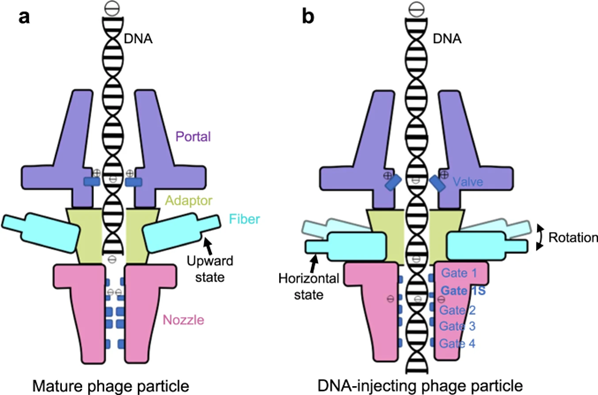
Proposed DNA gating model of P-SCSP1u during the DNA-injecting stage of infection.
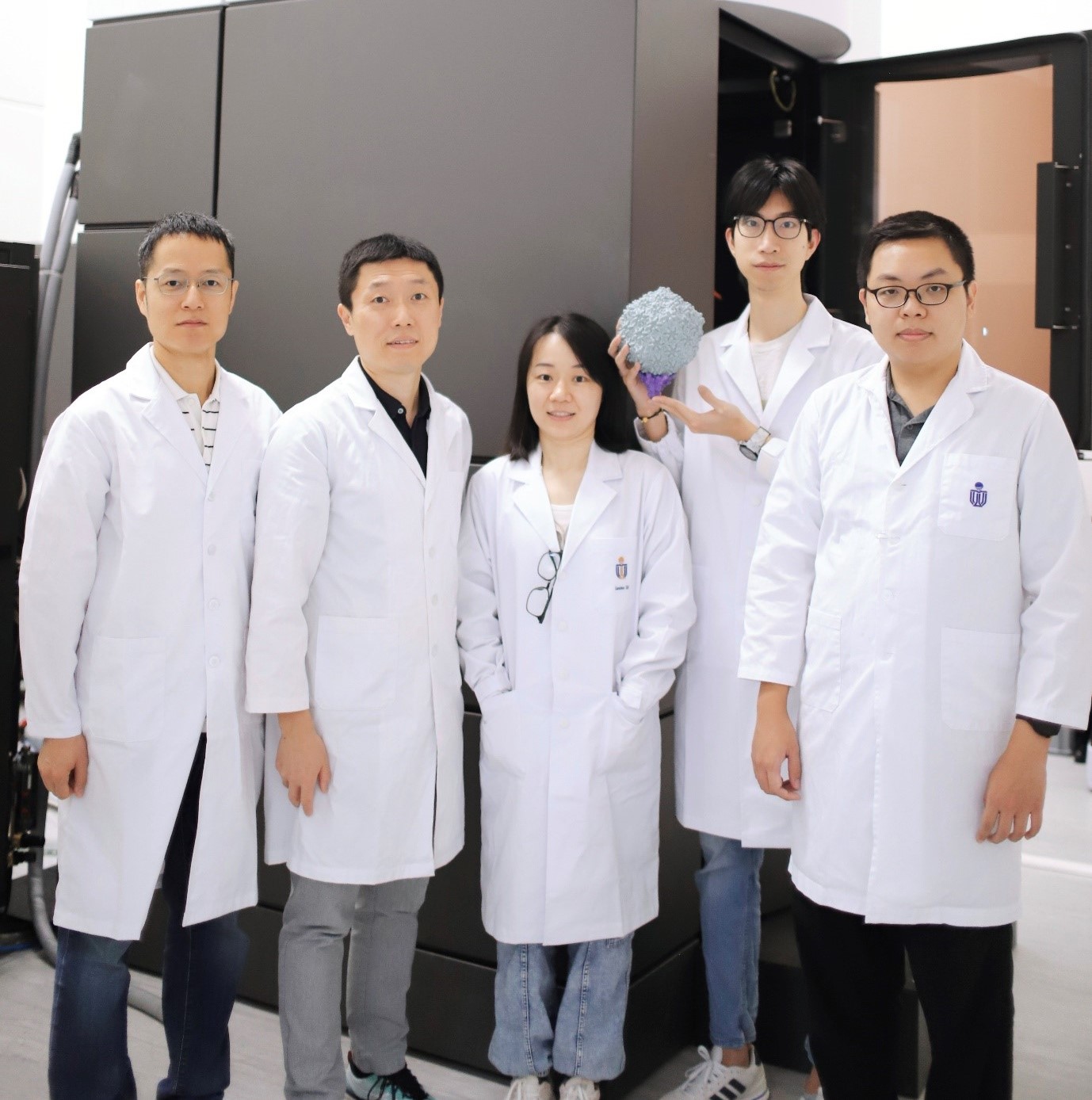
Photo of the research team, featuring Prof. DANG Shangyu, Prof. ZENG Qinglu, Dr. CAI Lanlan, Mr. LIU Hang (With the 3D printed cyanophage structure in his hands), and Mr. XIAO Shiwei, from left to right.
Scientific Breakthroughs & Discoveries

HKUST Elucidates How IGF2’s Secretory Pathway…
A research team led by the Hong Kong University of Science and Technology (HKUST) recently revealed how TMED10, a type of transmembrane protein, regulates muscle stem cell differentiation through mediating the secretion of insulin-like growth factor 2 (IGF2). This provides potential therapeutic strategies to downregulate IGF2 signaling by inhibiting its secretion.

HKUST Scientists Unveil Promising Target For…
A research team led by Prof. Nancy IP, the President and The Morningside Professor of Life Science at The Hong Kong University of Science and Technology (HKUST), and the Director of the Hong Kong Center for Neurodegenerative Diseases (HKCeND), has identified VCAM1, a cell surface protein found on immune cells of the brain, as a therapeutic target for Alzheimer’s disease (AD), paving the way for developing novel therapeutics to combat this debilitating condition.